Fatty Acids: Branched-Chain
 Branched-chain
fatty acids are common constituents of the lipids of bacteria and to a much lesser extent of animals,
although they are rarely found other than as surface lipids of higher plants.
Normally, the fatty acyl chain is saturated, and the branch is a methyl group, but unsaturated branched-chain fatty acids are found
in marine animals, while branches other than methyl may be present in microbial lipids.
The most common of these are mono-methyl-branched, but di- and poly-methyl-branched fatty acids (isoprenoid) are
known, while the mycolic acids from Mycobacteria have more complex structures.
In bacteria, their main function in membranes may be to increase the fluidity and lower the phase transition temperature of the lipid components as an alternative to the use of unsaturated fatty acids. As they have mainly saturated aliphatic chains, branched-chain fatty acids are not vulnerable to attack by reactive oxygen species, and this may be an explanation for their occurrence on surfaces exposed to oxygen flux, such as skin and tear films or leaf waxes. The following is an introduction to the structures, natural compositions and biosynthesis of these fatty acids, and it is not intended to be comprehensive.
1. Saturated iso- and anteiso-Methyl-Branched Fatty Acids
iso-Methyl branched fatty acids have the branch point on the penultimate carbon (one from the end or ω-1), while anteiso-methyl-branched fatty acids have the branch point on the ante-penultimate carbon atom (two from the end or ω-2) as illustrated. In the latter, the methyl group has the (S)‑configuration in general, reflecting its biosynthetic origin, but a small proportion of the (R)-enantiomer has been detected in some soil bacteria.

Fatty acids with structures of this type and with 10 to more than 30 carbons in the acyl chain are found in nature, but those most often encountered have 14 to 18 carbons in the chain (not counting the additional carbon in the methyl group). They are common constituents of bacteria, but they are rarely detected in other microorganisms. Via the food chain and commensal bacteria, they can be found in animal tissues, especially those of marine animals and ruminants (dairy products, beef and mutton) and these are a further source for the human diet, although they can be synthesised to some extent in animal tissues per se. In bacteria, their content and composition can often be used as taxonomic markers, and in the genus Bacillus, for example, some species have fatty acids with the iso-structure only, while others have the anteiso-structure.
Bacteria: These fatty acids are synthesised in bacteria via the conventional mechanisms for the synthesis of saturated fatty acids by FAS II (see the appropriate web page), except that the nature of the primer molecules differ. Thus, instead of acetyl-CoA, 2‑methylpropanyl-coA (derived from the amino acid valine) is the primer for the biosynthesis of iso-branched fatty acids with an even number of carbon atoms (odd-numbered chain), while 3‑methylbutyryl-CoA (derived from leucine) is the primer for iso-fatty acids with an odd number of carbon atoms (even-numbered chain). 2-Methyl-butyryl-CoA (derived from isoleucine) is the primer for anteiso-fatty acids to produce fatty acids with an odd number of carbon atoms (even-numbered chain). The first step is the conversion of each branched-chain amino acid to its corresponding α-keto acid by mitochondrial branched-chain amino acid transaminases, and this is followed by oxidative decarboxylation with the rate-limiting enzyme, branched-chain α-ketoacid dehydrogenase (BCKDC), which produces each of the primer-CoA esters. However, in Staphylococcus aureus, an ATP-dependent acyl-CoA synthetase has been characterized that is selective for 2-methylbutyric or isobutyric acids and is essential for branched-chain fatty acid synthesis.
 |
| Figure 1. Biosynthesis of iso- and anteiso-methyl-branched fatty acids. |
The committed step in the initiation of fatty acid synthesis is achieved by β-ketoacyl acyl carrier protein synthase (KAS III or FabH), which catalyses condensation of the primer CoA ester and malonyl-ACP, and the specificity of this enzyme determines whether FAS II can utilize branched precursors. While that in Escherichia coli cannot use them, Bacillus subtilis has two FabH enzymes, which both prefer branched-chain substrates. Determination of the crystal structure of KAS III in the facultative anaerobe Propionibacterium acnes has shown that the enzyme has a cavity at the catalytic site with a unique shape that favours branched-chain CoA precursors.
It is more common to find iso-methyl fatty acids with an even number of carbons in total, although the chain-length is odd-numbered, while the opposite is true for the anteiso-forms. Note: this sometimes leads to confusion in the informal nomenclatures that may be used in scientific publications. For example, 13-methyl-tetradecanoate acid is sometimes abbreviated to iso-methyl-14:0 and sometimes iso‑methyl-15:0; I prefer the former as it better reflects the systematic name, although many microbiologists tend to favour the latter. Because of the alternative route for iso-methyl formation and of subsequent alpha-oxidation processes, both odd- and even-numbered iso-methyl and anteiso-methyl acids can be present in tissues.
Under stress, bacteria maintain membrane fluidity through a mechanism termed "homeoviscous adaptation”, which may be achieved by synthesis of membrane lipids de novo or by acyl chain remodelling of the fatty acids of the existing membrane lipids. Methyl branching tends to decrease the thickness of lipid bilayers while lowering chain ordering to enhance the fluidity of membranes through the formation of kinks at the branching point. During cold stress, the proportion of anteiso-branched fatty acids of many bacteria increases because anteiso branching is more effective at fluidizing a membrane than iso branching, and this is crucial for the survival of such cold tolerant pathogens as Listeria monocytogenes. Thermophilic species, in contrast, have higher contents of iso-branched fatty acids that are often of a longer than average chain length. Other bacteria attain the same end by varying the proportion of unsaturated fatty acids in their membrane lipids.
Animals: In animal tissues, the biosynthesis of these fatty acids de novo is normally a minor process and involves a similar mechanism to the above with adipose tissue (but not liver or brain) as a significant source. Monomethyl branched-chain fatty acids are rapidly synthesised and then catabolized in brown adipose tissue especially by ACOX2 in peroxisomes to support energy dissipation. Catabolism of branched-chain amino acids in mitochondria generates the precursors, which are exported to the cytosol by adipose-specific expression of carnitine acetyltransferase. The fatty acid synthase then has sufficient flexibility to generate some long-chain monomethyl-branched fatty acids.
 Very-long-chain fatty acids
of this type (>C21) are synthesised from long-chain precursors by fatty acid elongases,
i.e., ELOVL1, ELOVL3 and ELOVL7, and this can be an important process in some tissues.
Lanolin, the waxy material produced as a protective coating for the fleece of sheep,
contains up to 45% of iso- and anteiso-branched fatty acids from C10 to C34 in chain-length
(see the web page on waxes).
One anteiso-branched fatty acid, 18-methyl-eicosanoic acid, constitutes up 60% of the total fatty acids
esterified directly to wool via thiol ester bonds, and it comprises 40% or more of the same lipid in all mammalian hairs examined to date.
A significant proportion of the wax and cholesterol esters secreted by the meibomian gland adjacent to the eye consist of
iso/anteiso-methyl-branched fatty acids, while ceramides in skin and liver can also contain such fatty acids.
In most mammalian tissues, branched-chain fatty acids rarely make up more that 1-2% of the total, and a high proportion is probably derived from commensal bacteria in the intestines or from consumption of such fatty acids in dairy products or meat from ruminant animals. Fish oils usually contain 1‑2% iso- and anteiso-fatty acids of chain-length C14 to C18, which are presumed to be derived from the marine food chain. Such fatty acids have been shown to have potentially beneficial effects in vitro by limiting the growth of breast cancer cells through inhibition of the synthesis of conventional fatty acids de novo, while a study with a human hepatoma cell line suggested that iso-C13:0 was especially effective by promoting apoptosis; there appear to be no studies of this phenomenon in vivo in humans. Among many other claimed benefits, they are reported to reduce intestinal inflammation in the human neonate.
The free-living nematode Caenorhabditis elegans, used as a model for research purposes, synthesises iso-methyl-tetradecanoic and iso-methyl-hexadecanoic acids de novo, and it has been shown to be absolutely dependent on these for its growth and development in the presence of elevated dietary glucose. Isovaleryl-CoA is elongated to iso-13:0 by the fatty acid synthase, then converted to iso-15:0 by a fatty acid elongase ELO-5 and further to iso-17:0 by ELO-5 and ELO-6. Acyl-CoA synthetases guide the incorporation of branched-chain acids into phospholipids and thus regulate their composition in the zygote stage. Disruption of the biosynthesis of these fatty acids by inhibiting the branched-chain α‑ketoacid dehydrogenase leads to early embryonic fatality, but supplementation with long-chain methyl-branched fatty acids corrects the problem.
Triacylglycerols and wax esters containing isovaleric (3-methylbutyric) acid and other branched-chain fatty acids are constituents of the blubber and melon (echo location) oils of the toothed whales and dolphins, and an alkyldiacylglycerol containing this acid occurs in rabbit harderian gland. Dolphins synthesise branched-chain fatty acids from leucine, whereas beaked whales use valine as the precursor, but enzymes that can elongate isovaleric acid are absent or limited in their activity.
Plants: In higher plants, branched-chain components are only rarely reported from the seeds or green tissues, but 14‑methylhexadecanoic occurs at a level of 0.5 to 1% in seed oils from the family Pinaceae, where it is a useful taxonomic marker, and anteiso-methyl-branched fatty acids have been characterized from the main tissues (non-wax) of Brussels sprouts. Plant surface waxes often have iso-/anteiso-methyl-branched fatty acids as major components, and in Arabidopsis, iso-branched wax biosynthesis utilizes isobutyric acid derived from valine as the precursor and requires an isobutyryl-CoA synthetase to initiate biosynthesis. Angelic acid or 2-methyl-2Z-butenoic acid is found linked to terpenoids in the roots of umbelliferous plants, such as Angelica archangelica.
Neo fatty acids: These can be considered as having a terminal tertiary butyl group or with two iso-methyl groups and have been found in some microorganisms, algae, plants and marine invertebrates. 13,13-Dimethyltetradecanoic acid or ‘neopalmitic’ acid illustrated is a minor component of bark and resins from conifers and other plants, and it has been found in the shell, chitin and chitosan of a crab.

2. Saturated Mid-Chain Methyl-Branched Fatty Acids
Bacterial fatty acids: 10R-Methyloctadecanoic or 'tuberculostearic' acid is a major component of the glycerophospholipids of Mycobacterium tuberculosis, where it controls lateral membrane partitioning, and in Corynebacterium and a few other bacteria. Its presence in bacterial cultures and sputum from patients is used in the diagnosis of tuberculosis. M. phlei contains a range of methyl-branched fatty acids, including 8- and 10‑methylhexadecanoate, 9‑methylheptadecanoate, 11‑methylnonadecanoate, 12‑methyleicosanoate, 14‑methyldocosanoate and 16‑methyltetracosanoate.

Many fatty acids with a single methyl branch of this kind have been isolated from other bacteria, and 10‑methylhexadecanoic and 11‑methyloctadecanoic acids are relatively common microbial constituents, while 12‑methylhexadecanoic acid and 14‑methyloctadecanoic acid are major components of the halotolerant bacterium Rubrobacter radiotolerans and of the aquatic bacterium Rhodococcus equi, and 6- and 9‑methyltetradecanoic acids are found in lichenized fungi. In mixed microbial populations such as those isolated from soil or other environmental samples, many different branched-chain fatty acids may be found.
Sponges and some other marine organisms contain methyl-branched fatty acids, but these are thought to be derived from microorganisms in their diet or that live in symbiosis with them. For example, as well as numerous iso- and anteiso-methyl-branched fatty acids, 10-methyl-16:0 to 20‑methyl‑26:0 are present in the lipids of the sponge Verongia aerophoba.
Biosynthesis of such branched-chain fatty acids involves methylation at the double bond of a monoenoic acid such as oleic or cis-vaccenic acid while esterified as a component of a phospholipid, with S-adenosylmethionine as the methyl donor. The resulting 10R‑methylene-octadecanoyl residue is reduced to the 10-methyl compound with NADPH as the cofactor; the 10-methylene-intermediate has been isolated from a Corynebacterium. A related mechanism is used for biosynthesis of cyclopropane fatty acids in bacteria, and the relevant enzyme from Mycobacterium smegmatis has been characterized as a cyclopropane-fatty-acyl-phospholipid synthase.
 |
| Figure 2. Enzymatic introduction of a methyl branch into an unsaturated fatty acid. |
Non-isoprenoid dimethyl-branched fatty acids are sometimes reported from bacteria, and 4,9‑dimethyl‑10:0, 4,10- and 4,11‑dimethyl-12:0, and 4,13-dimethyl-14:0 acids with 2,13- and 2,12-dimethyl-14:0 acids were identified in a halophilic Bacillus sp., while multi-branched fatty acids with the methyl branches in positions 2, 4, 6 and 8 are present in certain Mycobacteria (see below). Although dimethyl fatty acids are occasionally reported from sponges, they are presumed to be derived from bacteria in the food chain or that are symbiotic, e.g., 9,13- and 10,13‑dimethyl-14:0, 8,10-dimethyl-16:0 and 3,13-dimethyl-14:0.
Dimethyl, dicarboxylic acids termed diabolic acids, were first reported from rumen bacteria, such as Butyrivibrio fibrisolvens, but related fatty acids are common in the genus Acidobacteria. Diabolic acid per se is 15,16‑dimethyltriacontanedioic acid, while iso‑diabolic acid is 13,16‑dimethyloctacosanedioic acid. Some Acidobacteria from soils produce the latter with further methyl substituents in positions 5 or 6 relative to the one or both carboxyl groups, e.g., 5,13,16-trimethyloctacosanedioic acid. Similar fatty acids occur in some plant waxes.

Diabolic acid is formed biosynthetically by a tail to tail joining of two molecules of palmitic acid with no loss of hydrogen from positions 14 and 16, while iso-diabolic acid is formed in the same way by coupling of two molecules of iso-methyl-15:0. The relevant enzymes (membrane-spanning lipid synthases) were characterized in Thermoanaerobacter ethanolicus, an organism considered to be at the "lipid divide" between bacteria and the Archaea. Each of the carboxyl groups can link to glycerol moieties as part of a highly complex lipid, sometimes as tetraesters, which can span a membrane bilayer, often together with analogous ether lipids for which they may be the biosynthetic precursors by the action of glycerol ester reductases. In Acidobacteria, it is reported that iso-diabolic acid is formed by tale-to-tale condensation of two molecules of iso-methyl-tetradecanoic acid while these are linked to glycerol and not in a separate reaction prior to esterification. The additional 5/6-methyl group is inserted into the esterified fatty acid as a separate biosynthetic step. As the resulting complex lipids are based upon 1,2‑dialkyl/acyl-sn-glycerols, they differ in the stereochemistry of the glycerol moiety from the comparable lipids in the Archaea. This type of structure may provide greater stability to membranes under adverse pH conditions or at higher temperatures.
Animal sources: Fats from ruminants such as sheep can contain higher proportions of branched-chain components, especially those fed carbohydrate-rich diets when up to 9% of the subcutaneous fat can comprise such fatty acids. Relatively high proportions of propionic acid (as opposed to acetic and butyric) are produced by rumen microorganisms under this dietary regime, and this metabolite is converted to methylmalonyl-CoA, presumably by the promiscuous reaction of acetyl-CoA carboxylase on propionyl-CoA; ethyl malonate can be produced in the same way from butyryl-CoA and incorporated into fatty acids. Methylmalonyl-CoA is then incorporated into fatty acids by the fatty acid synthase (FAS I) at some point in the cycle of reactions. A consequence of this mechanism is that the methyl groups are all in the even-numbered positions, and they are distributed randomly in fatty acids of varying chain-lengths. In fact, more than 120 different mono-, di- and tri-methyl fatty acids (and some ethyl-branched components) have been identified in sheep fats. In other animals, the enzyme ECHDC1 has a repair function that serves to prevent the formation of the branched-chain intermediates.
 |
| Figure 3. Biosynthesis of branched-chain fatty acids in ruminant animals. |
Some such methyl-branched fatty acids occur in a few other disparate tissues in the animal kingdom. Perhaps the best known is the uropygial (preen) gland of birds, which secretes a waxy material that serves as waterproofing for the feathers, while providing olfactory cues and having antibiotic properties. While the precise composition of this varies among species, all are characterized by high concentrations of branched-chain fatty acids with alcohols and alkane-diols as esters. Most often the branch is a methyl group, but ethyl and propyl branches are known, and di-, tri- and tetramethyl-branched fatty acids are usually present. The positions of these and the chain-lengths of the various components cover a wide range, but for the monomethyl fatty acids, the branch-points are generally in positions 2 to 6. A common pattern is to find series such as 2,4-, 2,6-, 2,8- and 4,6-dimethyl and others, with 2,4,6-, 2,4,8- and 2,6,8-trimethyl, and 2,4,6,8-tetramethyl fatty acids in this wax, and in some birds, these can comprise 90% of the total fatty acids, but the preen gland of the barn owl contains 3‑methyl- and 3,5-, 3,7-, 3,9-, 3,11-, 3,13- and 3,15‑dimethyl-branched fatty acids. To illustrate the complexity, the composition of the fatty acids in the uropygial gland of the fulmar is listed in Table 1.
Table 1. Branched-chain components in the preen gland of the fulmar (wt% of the total). |
||
| Position | Chain-length | Amount (%) |
|---|---|---|
| 2- | C8 | 0.4 |
| 3- | C7 to C12 | 53.3 |
| 4- | C7 to C12 | 22.6 |
| 6- | C10 to C12 | 4.0 |
| 2,4-/2,6- | C8 to C10 | 6.5 |
| 3,7- | C9 to C11 | 8.3 |
| 4,6-/4,8- | C10 | 0.4 |
| Jacob, J. and Zeman, A. Z. Naturforsch., 26b, 33 (1971); DOI. | ||
Much remains to be learned of the mechanism of biosynthesis of these fatty acids, but again a high proportion at least is produced via methylmalonyl-CoA instead of malonyl-CoA for chain-elongation and insertion of the methyl group. Wax ester synthases have been identified.
A further interesting example is Vernix caseosa, the waxy skin secretion that covers new-born babies.
As well as a high proportion of iso- and anteiso-methyl-branched fatty acids, this contains approximately 10% of components
from C11 to C18 in chain-length with methyl groups in the even-numbered positions from 2 to 12 (more than 40 different
isomers), which are once more presumably synthesised using methylmalonate as a substrate.
These fatty acids have been detected in the intestines of the new-born, where they originate in lipid-laden vernix caseosa particles suspended
in amniotic fluid;  there is a suggestion that they may inhibit microbial attack.
A lactone derivative of a tetramethyl fatty acid hydroxylated on the carbon 3, i.e., with a highly strained four-membered lactone ring and known as ‘vittatalactone’ ((3R,4R)-3-methyl-4-(1,3,5,7-tetramethyloctyl)oxetan-2-one), is a pheromone emitted by the male striped cucumber beetles, Acalymma vittatum, which damage cucurbit crops in the U.S.A.
3. Isoprenoid Fatty Acids
Several isoprenoid fatty acids that are derived from the metabolism of phytol (3,7,11,15-tetramethylhexadec-2E-en‑1‑ol), the aliphatic alcohol moiety of chlorophyll, occur naturally in animal tissues. These range from 2,6-dimethylheptanoic to 5,9,13,17-tetramethyloctadecanoic acids, but those encountered most often are 3,7,11,15-tetramethylhexadecanoic (phytanic) and 2,6,10,14-tetramethylpentadecanoic (pristanic) acids. 4,8,12‑Trimethyltridecanoic acid is common in fish and other marine organisms. Phytanic acid occurs in tissues as a racemic mixture of (3R,7R,11R,15)- and (3S,7R,11R,15)-isomers.
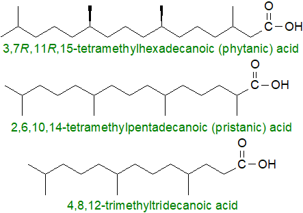
Normally, these fatty acids occur at low levels only in tissues, with the concentrations being highest in herbivores. Phytanic acid is found at levels of up to 1% normally in milk fat and adipose tissue from cows, but much higher concentrations can occur on occasion, and up to 20% of the fatty acids in the triacylglycerols of bovine plasma can consist of this acid because the methyl-branch in position 3 of the chain inhibits the action of the enzyme lipoprotein lipase, which clears triacylglycerols from plasma.
Dietary chlorophyll cannot be hydrolysed to phytol in the digestive system of humans, but rumen microorganisms can accomplish this before biohydrogenation of phytol to dihydrophytol occurs, followed by oxidation to phytanal and then further to phytanic acid. In the tissues of non-ruminant animals and some marine bacteria, phytanic acid is formed by oxidation of phytol first to phytenal and then to phytenic acid (with a double bond in position 2 and only encountered in tissues under artificial feeding conditions), followed by reduction. Most of the phytanic acid in the tissues of humans is ingested via meat and dairy products, although some is obtained from free phytol and phytol esters of fatty acids in certain vegetables, e.g., bell peppers and salad rocket. The shorter-chain isoprenoid fatty acids sometimes detected in tissues are formed from this by sequential α- and/or β‑oxidation reactions. Presumably, phytanic acid is formed in an analogous manner in fish and can thence enter the human food chain.
Because of the presence of the 3-methyl group, degradation of phytanic acid by β-oxidation cannot be initiated in animal tissues. Rather, phytanic acid is oxidized first by α-oxidation in peroxisomes, yielding pristanic acid, which can then be subjected to three cycles of β‑oxidation with 4,8‑dimethylnonanoyl-CoA as the end-product for transportation to mitochondria where full oxidation takes place. Some omega-oxidation can occur with 3-methyladipic acid as an end-product. In humans, several inborn metabolic errors in the degradation of phytanic and pristanic acids have been described that lead to an accumulation of these acids in tissues and body fluids. There are various clinical expressions of such disorders, some causing fatality, and the best known of these is Refsum’s disease, a rare genetic syndrome, in which defects in one or other steps in the α-oxidation system, but mainly in the enzyme phytanoyl-CoA 2‑hydroxylase, lead to the accumulation of phytanic acid in tissues and to clinical symptoms.
In contrast, there are suggestions that phytanic and pristanic acids may have some beneficial influences towards health that include protection against the metabolic syndrome, induction of the differentiation of brown adipocytes, regulation of aspects of glucose and retinoic acid metabolism, and inhibition of breast, colon and other cancers. They are signalling molecules that regulate the expression of those genes that affect the catabolism of lipids. By binding to a liver-type fatty acid binding protein, they are transported to the nucleus where they exert their effects by interacting with peroxisome proliferator-activated receptor alpha (PPARα), which induces the transcription of enzymes for fatty acid degradation by β- and ω-oxidation. In a sense, they are regulators of their own catabolism.
Retinoic acid, an isoprenoid fatty acid derived from retinol (vitamin A), is a regulator of genes for cell growth and differentiation via transport proteins and nuclear receptors, but it has its own web page here. In contrast to phytanic acid, it is not found esterified to mainstream lipids in tissues. Dolichoic acids, derived from dolichols and containing 14 to 20 isoprene units, have been found in lipid extracts of human brain.
4. Unsaturated Methyl-Branched Fatty Acids
Small amounts of monounsaturated methyl branched-chain fatty acids have been detected in the lipids of the human sebaceous gland, i.e., iso-methyl-6Z-16:1 and anteiso-methyl-6Z-17:1, as 4% and 0.8%, respectively, of the skin surface lipids, with human fatty acid desaturase 2 (FADS2), which produces sapienic acid (6Z‑16:1) in skin, as the enzyme responsible for introduction of the double bond. Similar fatty acids with iso-/anteiso-methyl groups detected in related marine organisms include 13‑methyltetradec-4-enoic, 14‑methylhexadec-6-enoic, 14‑methylpentadec-6-enoic and 17‑methyloctadec-8-enoic acids. It is possible that the primary origin of these fatty acids is in bacteria, as such species as Bacillus cereus, B. megaterium and Desulfovibrio desulfuricans contain many comparable fatty acids. On occasion, the branch is more central in the aliphatic chain, and one of the first acids of this type to be described was 7‑methyl-7-hexadecenoic acid from lipids of the ocean sunfish, Mola mola, but now known to be more widespread in fish from warmer seas, while 7-methyl-6- and 7‑methyl-8-hexadecenoic acids were later found in sponges. (Z)‑13‑Methyltetra-4-decenoic acid ((Z)‑4c‑14:1), isolated from a marine sediment bacterium Olleya marilimosa, has antibacterial properties against Gram-positive bacteria.

Many different demospongic acids, i.e., with bis-methylene-interrupted double bonds (usually in the 5,9-positions), together with some monoenes have been described with iso- and anteiso-methyl branches (see our web page dealing with demospongic acids), and several related fatty acids have been described with the methyl group in more central positions, e.g., 17-methyl-5,9-24:2, 21-methyl-5,9-26:2 and 22-methyl-5,9-28:2. Unusual multibranched polyunsaturated and very-long-chain fatty acids have been located in slime moulds and freshwater sponges from Israel, including 7,9,13,17-tetramethyl-7S,14S-dihydroxy-2E,4E,8E,10E,12E,16-octadecahexaenoic acid from seven different myxomycetes.
Branched-chain unsaturated fatty acids are not common in plants, but small amounts of 16-methyl-cis-9-octadecenoic and 16‑methyl-cis-9,cis-12-octadecadienoic acids have been found in wood and seeds of certain Gymnosperms.
5. Mycolic and Related Fatty Acids from Mycobacteria
The genus Mycobacterium consists of numerous highly unusual aerobic species, some of which are pathogens responsible for such diseases as tuberculosis and leprosy. They have a unique cell wall structure and contain many lipid classes found nowhere else in nature (summarized below). Of these, the mycolic acids are β‑hydroxy-α-alkyl branched fatty acids of high molecular weight, which are structurally diverse with respect to their functional groups and carbon chain length, depending on the mycobacterial species and strain. In the genus Mycobacteria, they can have 60 to 90 carbons, and they are major components of the lipid components of the cell walls and of those of the related genera Nocardia, Rhodococcus and Corynebacterium. Those of M. tuberculosis are of particular interest because of the pathogenicity of this organism (see below).
The main structural and characteristic unit of mycolic acids is termed a "meromycolic (or mero) chain", and it can contain a variety of substituents, including double bonds of both the cis- and trans-configurations (but when the latter, they also possess an adjacent methyl branch) and cyclopropane rings, which can likewise be of the cis- and trans-configurations. Further complexity comes from hydroxy, methoxy-, epoxy- and keto substituents of defined stereochemistry, which are adjacent to a methyl branch normally. The secondary or alpha-branched unit linked to position 2 of the merochain commonly consists of a C22 to C26 unsubstituted alkyl moiety, while position 3 almost invariably has a hydroxyl group. The 2R,3R-stereochemistry of the substituents at positions 2 and 3 is conserved in all genera and is crucial for T-cell recognition. Mycolic acids are major components of the cell wall linked to lipopolysaccharides, but they also occur in free form or esterified to trehalose or phospholipids.
The least polar mycolic acids, often termed alpha-mycolic acids, contain 74-80 carbon atoms and generally two double bonds (of the cis- or trans-configuration) or two cis-cyclopropyl groups located in the meromycolic chain, and while some may contain further double bonds, e.g., in M. tuberculosis, they have no oxygenated substituents other than the hydroxyl in position 3. Cyclopropyl rings in the alpha-mycolates tend to have the cis-configuration. The alpha'-mycolic acids have 60-62 carbon atoms and one cis double bond. In M. tuberculosis, the oxygenated mycolic acids are 4-6 carbon atoms longer than the non-polar equivalents, and methoxy forms are the second most abundant class after the alpha-mycolates, followed by the keto-mycolates. The genus Segniliparus contains a distinctive series of mycolic acids, including the longest known (C42-C100) in which the alpha-mycolates lack the hydroxyl group in position 3. Some representative structures are illustrated.
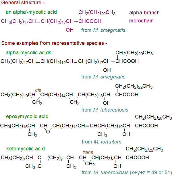 |
| Figure 4. Structures of some typical mycolic acids. |
The β-hydroxyl group in mycolic acids modulates both the phase transition temperature and the molecular packing within the membrane, while cyclopropane rings contribute to the structural integrity of the cell wall complex and are protective against oxidative stress; trans-cyclopropyl mycolates assist in maintaining cell wall viscosity under diverse physiological conditions. When the structures of the mycolic acids are changed experimentally, for example by trans-cyclopropanation in a distal position, there is a loss of virulence, but if this occurs close to the carboxyl group there is an opposite effect. It is evident that oxygenated moieties are required for maximum toxicity.
Biosynthesis: The two component parts of mycolic acids are each synthesised by different enzyme systems in the microorganisms before they are condensed to form a typical mycolic acid. In M. tuberculosis, a fatty acid synthase I (FAS I) of the animal type has a bimodal product profile and provides C24 to C26 fatty acids, corresponding to the α-branch in mycolic acid synthesis, and C16 to C18-acyl-CoAs to a fatty acid synthase II system (FAS II, plant or prokaryotic) for chain-elongation via iterative elongation cycles into the very-long-chain fatty acids, the meromycolic acids (C36 to C72). FAS I is the only system capable of synthesising fatty acids de novo (see our web pages on the biosynthesis of saturated fatty acids for further details of fatty acid synthases). In addition to mycolic acid production, FAS I products can be transferred to polyketide synthases to be utilized for the synthesis of other lipids, including phenolphthiocerols and mycocerosic acids.
 Cis
double bonds are introduced at two locations on a growing meroacid chain (two aerobic desaturases have been identified designated DesA1 and DesA2)
to yield three different forms of cis,cis-diunsaturated fatty acyl intermediates, which can then be converted enzymatically to methyl,
cyclopropane, methoxy- and/or keto-meroacids.
Ten related cyclopropane mycolic acid synthases are also S‑adenosylmethionine-dependent methyltransferases, each with strong
substrate and reaction specificities, and they can produce methyl-alcohol and other groups as well as cyclopropanes from double bonds.
For example, PcaA is a cyclopropane synthase that adds the proximal cis-cyclopropanes of cis/cis-meromycolates, a reaction
that is of special relevance in the early stages of infection; the enzyme is structurally different from the
E. coli analogue.
Finally, the mature meroacids and a C26‑S-acyl carrier protein enter into a Claisen condensation
catalysed by a polyketide synthase to yield the mycolic acids.
Transport of the mycolates to either the cell envelope or for attachment to arabinogalactans or to trehalose dimycolates via ester bonds
occurs in the form of trehalose monomycolates, although they are also present in the cell wall
as glucose monomycolate, glycerol monomycolate and in unesterified form.
Certain T cells can recognize mycolic acids and play a protective role during infection by M. tuberculosis, and there is hope that new vaccines will recognize both lipid and protein antigens. Recent findings that mycolic acids from M. tuberculosis and cholesterol interact with each other and bind to similar molecules are leading to a new understanding of host-pathogen interactions, which may lead to better control of the disease. Epoxyl groups can be converted to diols by specific hydrolases, possibly as a response to oxidative stress.
Analysis: Structural analysis of such complex fatty acids is much more difficult than with conventional fatty acids and usually a battery of chromatographic and spectroscopic techniques must be employed. One useful reaction uses pyrolysis to yield an alpha- or mero-fatty acid containing all the substituent groups and a mero-aldehyde, which can be analysed separately by mass spectrometry.
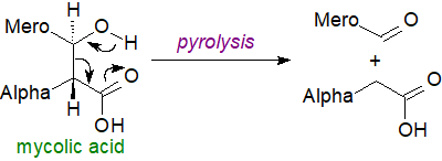 |
| Figure 5. Degradation of mycolic acids by pyrolysis for analytical purposes. |
Phthioceranic and mycobacteric acids: Mycobacteria contain some multi-methyl-branched fatty acids (C14 to C32 in chain length), often with the methyl branches in positions 2, 4, 6 and 8, i.e., 1,3-branched, and sometimes with hydroxyl groups in position 3. The phthioceranic acids are hepta- or octamethyl fatty acids, some of which are hydroxylated (hydroxyphthioceranic acid), and related forms include mycoceranic (2,4,6‑trimethyloctacosanoic), mycolipenic (2,4,6-trimethyl-trans-2-tetracosenoic), mycocerosic (2,4,6,8-tetramethyl-dotriacontanoic), and mycolipanolic (2R,4S,6S-trimethyl-3R-hydroxy-tetracosanoic) acids.
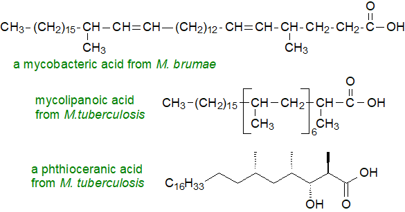 |
| Figure 6. Structures of some other branched chain acids from Mycobacteria. |
These occur together with long-chain (C36 to C47) fatty acids termed 'mycobacteric' acids, which are related structurally to mycolic acids and contain cyclopropyl rings, double bonds (trans and cis) and/or oxygenated substituents. Indeed, the latter are formed from the mycolic acids by a cleavage between carbons 3 and 4 by a Baeyer-Villiger-like reaction. Mycobacteric acids are present esterified to one of the three hydroxyl groups of triacylglycerols.
Cell wall of M. tuberculosis: More than a million people die from tuberculosis every year. A key factor in the persistence of M. tuberculosis infections is its unique and complex cell wall, a high proportion of which consists of complex and often unique lipids, many of which are linked to mycolic acids. These confer extreme hydrophobicity to the outer surface, resist degradation by host enzymes, including those of the immune system, and by antibiotics, and are associated with its pathogenicity. It has a dual membrane structure, illustrated schematically below, that is neither fully Gram-negative nor Gram-positive.
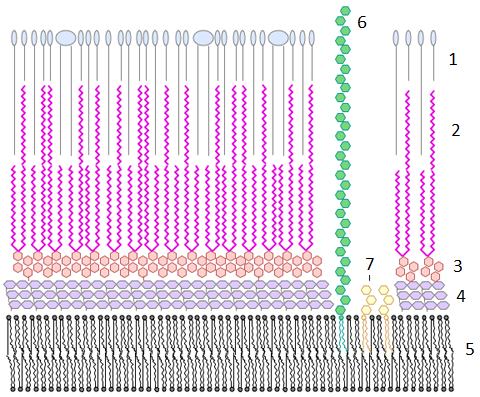
Figure 7. Schematic diagram of the cell wall of M. tuberculosis (not to scale). 1, Outer layer of waxy lipids; 2, mycolic acids linked to - 3, polysaccharides (arabinogalactan); 4, peptidoglycan layer (periplasm); 5, inner or plasma membrane; 6, lipoarabinomannan; 7, phosphatidylinositol mannoside. Reproduced with thanks under a Wikipedia creative commons license from Y tambe.
The cell-wall is capped by a loosely bound structure termed the capsule (not illustrated), which consists primarily of polysaccharides and peptides. The outermost layer of the outer membrane or mycomembrane facing the environment is highly hydrophobic and is composed largely of waxy lipids, such as di-, tri- and penta-acyl-trehaloses, dimycocerosates of phthiocerols and phthiodiolones and sulfated trehalose glycolipids, with some secreted proteins; these are not attached covalently to the cell wall. Mycolic acids are major constituents and structural components of the inner leaflet of the outer membrane, where they exist both in unesterified form and esterified to distinctive lipids, e.g., trehalose mono- and di-mycolates and lipopolysaccharides such as the underlying arabinogalactan of the arabinogalactan–peptidoglycan matrix. In Corynebacteria, they are also present O‑linked to proteins. The mycolic acid chains pack together in parallel arrays perpendicular to the surface of the cell wall, and they are stabilized by hydrogen bonds between the β-hydroxyl groups. In esterified form, keto-mycolic acids probably adopt a singular tight 'W' conformation. Mycolic acids are responsible for many of the distinctive properties of the membranes, including a low permeability to hydrophobic compounds in general and β‑lactam antibiotics in particular, as well as resistance to dehydration. Substantial amounts of free mycolic acids are present in the extracellular matrix of mycobacteria grown as biofilms, and indeed they are required for this type of growth.
In the mycobacterial inner membrane, there are conventional phospholipids such as cardiolipin, phosphatidylethanolamine, phosphatidylglycerol and phosphatidylinositol with the phosphatidylinositol mannosides; tuberculostearic acid (see part 2 above) is a major component of these. Mycolic acids have been characterized in position sn-2 of phosphatidylglycerol of Corynebacterium glutamicum but not in pathogenic C. striatum. The inner leaflet of this contains a high proportion of tri- and tetra-acylated phosphatidylinositol dimannoside, while the outer leaflet contains mainly tri-/tetra-acylated phosphatidylinositol hexamannosides. In the central periplasmic region, where enzymatic reactions occur, lipomannans and lipoarabinomannans extend outwards from lipid anchors in the inner membrane. Triacylglycerols present in the cell wall as well as the cytoplasm may be required for energy production when the organism is dormant in a host animal.
For practical reasons on this website, many of the unique lipids in cell walls of Mycobacteria are discussed at greater length with structurally analogous lipids on other web pages; these include phosphatidylinositol mannosides, lipomannans, phenolphthiocerol and phthiocerol dimycocerosates, trehalose esters, which include di- to penta-acyltrehaloses and sulfatides, glucose monomycolates, mannosyl-phosphomycoketide, gentiobiosyl-diacylglycerols, lysyl-diacylglycerols, (glyco)-peptidolipids, 6-O-methylglucose–containing lipopolysaccharides, isonitrile-diacyldipeptides (kupyaphores), phleic acids and triacylglycerols (not to forget tuberculostearic and related fatty acids discussed above).
Recommended Reading
- Batt, S.M., Minnikin, D.E. and Besra, G.S. The thick waxy coat of mycobacteria, a protective layer against antibiotics and the host's immune system. Biochem. J., 477, 1983-2006 (2020); DOI.
- Campagna, S., Mardon, J., Celerier, A. and Bonadonna, F. Potential semiochemical molecules from birds: A practical and comprehensive compilation of the last 20 years studies. Chem Senses, 37, 3–25 (2012); DOI.
- Chen, Y.F., Yang, H., Zheng, F.F., Wu, R.J., Zhang, C.L., Naafs, B.D.A., Pancost, R.D. and Zeng, Z.R. Temperature-dependent modulation of the methylation degree of (tetra) ester-linked membrane-spanning lipids in an Acidobacterium. Geochim. Cosmochim. Acta, 401, 190-203 (2025); DOI.
- Dembitsky, V.M. Natural neo acids and neo alkanes: their analogs and derivatives. Lipids, 41, 309-340 (2006); DOI.
- Gozdzik, P., Magkos, F., Sledzinski, T. and Mika, A. Monomethyl branched-chain fatty acids: Health effects and biological mechanisms. Prog. Lipid Res., 90, 101226 (2023); DOI.
- Hanhoff, T., Benjamin, S., Börchers, T. and Spener, F. Branched-chain fatty acids as activators of peroxisome receptors. Eur. J. Lipid Sci. Technol., 107, 716-729 (2005); DOI.
- Hauff, S. and Vetter, W. Exploring the fatty acids of vernix caseosa in form of their methyl esters by off-line coupling of non-aqueous reversed phase high performance liquid chromatography and gas chromatography coupled to mass spectrometry. J. Chromatogr. A, 1217, 8270-8278 (2010); DOI.
- Jackson, M., Stevens, C.M., Zhang, L., Zgurskaya, H.I. and Niederweis, M. Transporters involved in the biogenesis and functionalization of the mycobacterial cell envelope. Chem. Rev., 121, 5124-5157 (2021); DOI.
- Jia, F., Cui, M., Than, M.T. and Han, M. Developmental defects of Caenorhabditis elegans lacking branched-chain α-ketoacid dehydrogenase are mainly caused by monomethyl branched-chain fatty acid deficiency. J. Biol. Chem., 291, 2967-2973 (2016); DOI.
- Koopman, H.N. Function and evolution of specialized endogenous lipids in toothed whales. J. Exp. Biol., 221, jeb161471 (2018); DOI.
- Lu, H.J., Wang, Z., Cao, B., Cong, F., Wang, X.G. and Wei, W. Dietary sources of branched-chain fatty acids and their biosynthesis, distribution, and nutritional properties. Food Chem., 431, 137158 (2024); DOI.
- Mao, S.Q. and others. Branched-long-chain monomethyl fatty acids: are they hidden gems? J. Agric. Food Chem., 71, 18674-18684 (2023); DOI.
- Prithviraj, M. and others. Tuberculostearic acid controls mycobacterial membrane compartmentalization. mBio, 14, e0339622 (2023); DOI.
- Rafal, S., Magdalena, D., Karol, M., Bartlomiej, S. and Konrad, K. Detection and accurate identification of Mycobacterium species by flow injection tandem mass spectrometry (FIA-MS/MS) analysis of mycolic acids. Sci. Rep., 15, 13118 (2025); DOI.
- Roca-Saavedra, P., Mariño-Lorenzo, P., Miranda, J.M., Porto-Arias, J.J., Lamas, A., Vazquez, B.I., Franco, C.M. and Cepeda, A. Phytanic acid consumption and human health, risks, benefits and future trends: A review. Food Chem., 221, 237-247 (2022); DOI.
- Sahonero-Canavesi, D.X. and 11 others. Disentangling the lipid divide: Identification of key enzymes for the biosynthesis of membrane-spanning and ether lipids in Bacteria. Sci. Adv., 8, eabq8652 (2022); DOI.
- Siliakus, M.F., van der Oost, J. and Kengen, S.W.M. Adaptations of archaeal and bacterial membranes to variations in temperature, pH and pressure. Extremophiles, 21, 651-670 (2017); DOI.
- Wallace, M. and others. Enzyme promiscuity drives branched-chain fatty acid synthesis in adipose tissues. Nature Chem. Biol., 14, 1021-1031 (2018); DOI.
For tutorials on mass spectral analysis of fatty acids - see our mass spectrometry pages.
 |
© Author: William W. Christie |  |
|
| Contact/credits/disclaimer | Updated: November 2025 | ||
© The LipidWeb is open access and fair use is encouraged but not text and data mining, AI training, and similar technologies.
