Mass Spectrometry of Dimethyloxazoline and Pyrrolidine Derivatives
Halogenated Fatty Acids
As with my other documents on mass spectrometry, this is a subjective account that details only those fatty acids relevant to this topic that have been encountered during our research activities and for which we have spectra available for illustration purposes. Regretfully, we recognize that this a rather incomplete account of this subject. Spectra of DMOX derivatives with pyrrolidides are described here, but those of methyl esters and 3‑pyridylcarbinol ('picolinyl') esters are discussed on separate web pages. Key diagnostic ions only are described as general features of these derivatives are described elsewhere on this website. Only a few of these spectra have been published elsewhere.
Despite having very different structures, dimethyloxazoline and pyrrolidide derivatives of a given fatty acid have identical molecular weights and fragment in a remarkably similar way in the mass spectrometer. For this reason, it is convenient to discuss both together here. DMOX derivatives tend to give more abundant diagnostic ions and have better gas chromatography properties. However, pyrrolidides give more informative spectra when the functional group is close to either end of the molecule.
DMOX Derivatives of Fluoro Fatty Acids
One natural seed oil contains several fatty acids with a fluorine atom on the terminal carbon, i.e., Dichapetalum toxicarium. When we re-investigated this oil using 3‑pyridylcarbinol ester and DMOX derivatives, we found several hitherto unknown minor fatty acid components in addition to obtaining novel mass spectrometric information (Christie et al., 1998). We have some mass spectra from several synthetic fluoro-fatty acids kindly donated by Dr David O'Hagan of St Andrews University, and details of some of these have been published (Hamilton and Christie, 2000). They provided important evidence for some mechanistic concepts, but the aim of these pages is simply to act as a practical guide.
As an example, the mass spectrum of the DMOX derivative of 18-fluoro-octadecanoate -

There is nothing in the spectrum to confirm the position of the fluorine atom (cf., the spectrum of the pyrrolidide derivatives below). After the small molecular ion, there is a gap of 15 amu to m/z = 340, representing loss of a methyl group, not of ‑CH2F. We now know that the explanation is a facile loss of a methyl group from the ring structure that confounds the picture (see Hamilton and Christie, 2000). In the high mass range, the first ion that lacks fluorine is at m/z = 294.
The mass spectrum of the DMOX derivatives of 18-fluoro-octadec-9-enoate -

The pattern is repeated in that a loss of 15 amu from the molecular ion to m/z = 338 confuses the picture at the terminal carbon. In the high mass range, the first ion that we can be sure lacks fluorine is at m/z = 292. The position of the double bond is determined as described in the section of this website on DMOX derivatives of monoenes.
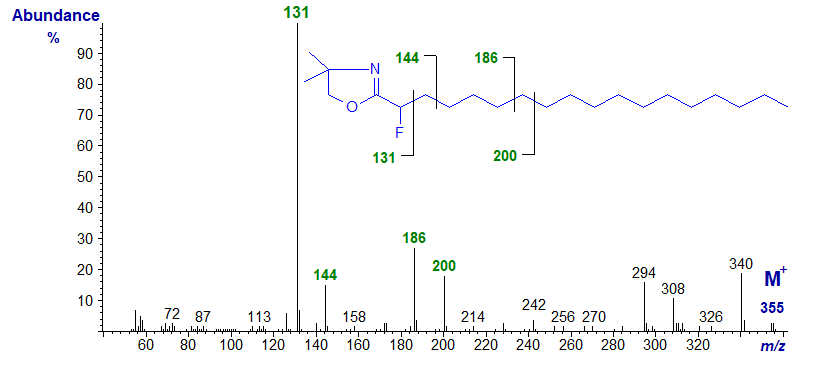
Mass spectrum of the DMOX derivative of 2-fluoro-octadecanoate (synthetic standard) -

The McLafferty ion at m/z = 131 has been shifted upwards by 18 amu (from 113) as expected, and most other ions are 18 amu higher in comparison to the non-fluorinated analogue. As in the spectrum of the corresponding methyl ester derivative, prominent ions at m/z = 186 and 200 in this instance indicate preferential cleavages between C-6 and C-7, and C-7 and C-8, respectively.
Further mass spectra of fluorinated fatty acids in the form of DMOX derivatives are available, but without interpretation, in the Archive Section of these web pages.
Pyrrolidine Derivatives of Fluoro Fatty Acids
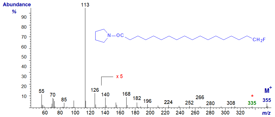
There is much less confusion in the identification of pyrrolidine derivatives when the fluorine atom is in the terminal position, as in spectrum of the pyrrolidine derivative of 18‑fluoro-octadecanoate -

The ions in the high mass range are less abundant than in the spectrum of the DMOX derivative (at least relative to the base ion), and it is necessary to magnify them to study the fragmentations. Following the molecular ion, there is a gap of 20 amu to m/z = 335 (loss of HF) for loss of −CH2F. From the spectrum, it could be argued that the fluorine atom is in either position 17 or 18, but at least this is a step forward. There is no confusing ion at [M−15]+ as with the DMOX derivative because there are no methyl groups on the ring.
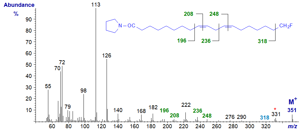
The mass spectrum of the pyrrolidine derivative of 18-fluoro-octadec-9,12-dienoate (linoleate) is -

Again the ions in the high mass range are less abundant than in the spectrum of the DMOX derivative, and there is no confusing ion at [M−15]+ as with the DMOX derivative. The diagnostic ions for the double bonds are clearly delineated (see the section of this website on Pyrrolidine derivatives of dienes). Following the molecular ion, there is a gap of 20 amu to m/z = 331 (loss of HF) and of 33 amu to m/z = 318 (very small) for loss of −CH2F, defining the structure.
Further mass spectra of fluorinated fatty acids in the form of pyrrolidine derivatives are available, but without interpretation, in the Archive Section of these web pages.
DMOX Derivatives of Bromo Fatty Acids
Brominated fatty acids have been found in many marine organisms, but we do not have access to these. While fluoro fatty acids were derivatized without difficulty, our first attempts to prepare derivatives of bromo fatty acids (produced by microbial fermentation of bromohydrocarbons) failed when derivatization procedures caused elimination of bromine with formation of ethoxy or methoxy fatty acids (see the section of this website on mass spectrometry of methyl esters of epoxy, furanoid and alkoxy fatty acids). Synthetic 2‑bromo-hexadecanoic acid was obtained from a commercial source and was successfully derivatized by our relatively mild two-step method (see the section of this website on Preparation of derivatives for mass spectrometry).
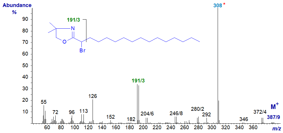
The mass spectrum of the DMOX derivative of 2-bromo-hexadecanoic acid -

The molecular ion is a small doublet at m/z = 387/9 (bromine exists as two isotopes in approximately equal amounts of atomic mass 79 and 81) and there is a small doublet at m/z = 372/4 for loss of a methyl group (probably from the ring system). Again, the key diagnostic feature that locates the bromine on carbon 2 is the fact that the McLafferty ion forms a doublet at m/z = 191/3. The base ion at m/z = 308 reflects loss of bromine from the molecule.
References
- Christie, W.W., Hamilton, J.T.G. and Harper, D.B. Mass spectrometry of fluorinated fatty acids in the seed oil of Dichapetalum toxicarium. Chem. Phys. Lipids, 97, 41-47 (1998); DOI.
- Hamilton, J.T.G. and Christie, W.W. Mechanisms for ion formation during the electron impact-mass spectrometry of picolinyl ester and 4,4-dimethyloxazoline derivatives of fatty acids. Chem. Phys. Lipids, 105, 93-104 (2000); DOI.
| © Author: William W. Christie |  |
|
| Updated: May 2025 | Contact/credits/disclaimer | |
© The LipidWeb is open access and fair use is encouraged but not text and data mining, AI training, and similar technologies.
